Department of Mechanical and Metallurgical Engineering, Institute for Biological and Medical Engineering.

Portada » Fibernet: Modeling heart muscle fibers with AI to treat arrhythmias

Department of Mechanical and Metallurgical Engineering, Institute for Biological and Medical Engineering
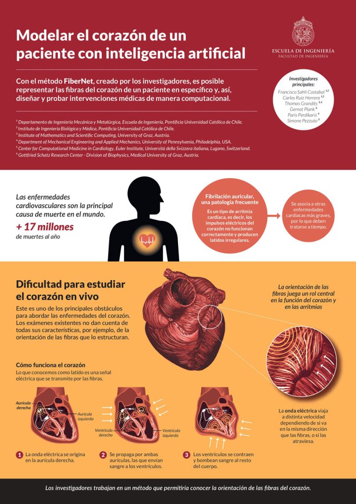
What we commonly refer to as “heartbeats” consist of a series of electrical signals that spread through the cardiac fibers, an arrange of muscle “threads” that extend across the entire surface of the heart, disposed in different directions. The orientation of these fibers varies from one person to another and, to date, we lack medical techniques or procedures that would allow us to visualize them in a living heart.
Why is it important to know the orientation? The arrangement of these fibers is a very relevant factor in the behavior of the electrical wave that generates every heartbeat: its velocity depends on whether it propagates along the same direction or if it crosses the fibers at some point. When a malfunction of the electrical impulses of the heart causes the beating to become irregular – a condition known as “cardiac arrhythmia” – the orientation of the fibers may be key to design an effective treatment and prevent the progression to more serious cardiac pathologies.
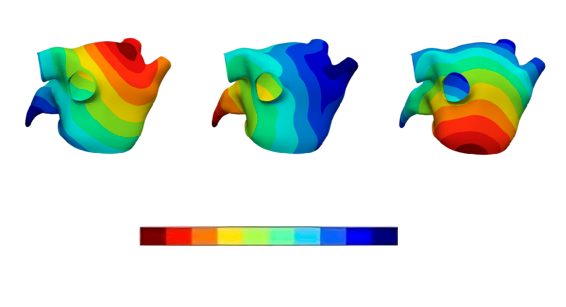
Professor Francisco Sahli and his research team leveraged artificial intelligence (AI) techniques to develop a method that enables mapping the entire network of heart fibers in a patient. This method was named ‘Fibernet’ and it can be broadly described as follows: a catheter is inserted into the atrium of the heart, carrying an electrode that captures the progression of electrical waves, which are then translated into a “cardiac activation map” illustrating the propagation of waves in time and space.

Cardiac activation map with colors indicating activation times.
This information is then processed using AI techniques collectively known as “machine learning”, where computational algorithms imitate certain elements of the learning process of our brains, but with a volume of information and complexity that surpasses by far what a person can normally grasp.
Thus, the cardiac activation maps obtained with the catheter are computationally processed through “artificial neural networks” (computational models that can process and learn from complex information). Since this information is not sufficient to calculate the orientation of the fibers, it is complemented with general physical laws that describe the way in which waves propagate and are therefore applicable to the electrical signals of the heart.
Department of Mechanical and Metallurgical Engineering, Institute for Biological and Medical Engineering

This technique used in artificial intelligence is called PINNs (physics-informed neural network) and consists of incorporating laws of physics to replace missing information.
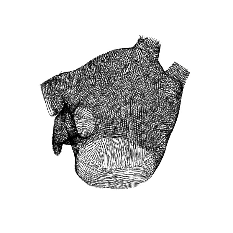
Combining these sources of information, the team was able to project the orientation of heart fibers with a precision never achieved before, which could lead to great advances in cardiology and medicine.

3D model of the heart obtained using Fibernet with the prediction of fiber orientations
For example, surgical treatments for cardiac arrhythmia, such as fiber ablation, could be tested digitally before intervening on the patient. In the ablation procedure, some points of the heart fibers are cauterized to reroute the irregular electrical signals causing arrhythmia episodes. With Fibernet, the exact point of the heart fiber needing ablation could be found using a virtual representation of the heart. This may lead to a higher success rate of the procedure and would be an important step towards a more personalized medical practice.

Department of Mechanical and Metallurgical Engineering, Institute for Biological and Medical Engineering.